Fluorescence Microscopy Digital Image Gallery
Transformed African Green Monkey Kidney Fibroblast Cells (COS-7 Line)
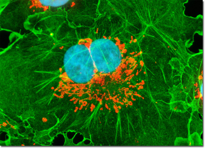
The culture of COS-7 fibroblasts illustrated in the digital image above was labeled for mitochondria with MitoTracker Red CMXRos and for the cytoskeletal filamentous actin network with Alexa Fluor 488 conjugated to phalloidin, a cyclic peptide derived from the toxic death cap fungus (Amanita phalloides). In addition, the fibroblasts were counterstained for DNA in the cell nucleus with Hoechst 33258. Images were recorded in grayscale with a Hamamatsu ORCA-AG camera system coupled to an Olympus BX-51 microscope equipped with bandpass emission fluorescence filter optical blocks provided by Semrock. During the processing stage, individual image channels were pseudocolored with RGB values corresponding to each of the fluorophore emission spectral profiles.