Fluorescence Microscopy Digital Image Gallery
Bovine Pulmonary Artery Endothelial Cells (BPAE Line)
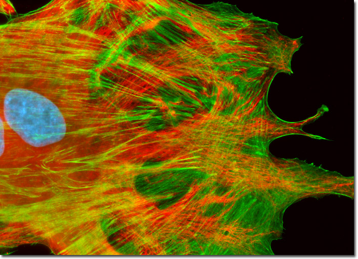
In order to label the intermediate filaments in the log phase adherent BPAE culture illustrated above, the fixed and permeabilized cells were blocked and treated with mouse anti-vimentin (porcine eye lens) primary antibodies followed by goat anti-mouse secondary antibodies (IgG) conjugated to Texas Red-X. Filamentous actin was visualized with phalloidin conjugated to Oregon Green 488, while the nuclei were stained with Hoechst 33258. Images were recorded in grayscale with a Hamamatsu ORCA-AG camera system coupled to an Olympus BX-51 microscope equipped with bandpass emission fluorescence filter optical blocks provided by Semrock. During the processing stage, individual image channels were pseudocolored with RGB values corresponding to each of the fluorophore emission spectral profiles.