Image Galleries
Featured Article
 Electron Multiplying Charge-Coupled Devices (EMCCDs)
By incorporating on-chip multiplication gain, the electron multiplying CCD achieves, in an all solid-state sensor, the single-photon detection sensitivity typical of intensified or electron-bombarded CCDs at much lower cost and without compromising the quantum efficiency and resolution characteristics of the conventional CCD structure.
Product Information
Digital Image Gallery
Fluorescence Microscopy Digital Image Gallery
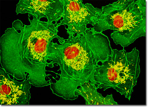
Transformed African Green Monkey Kidney Fibroblast Cells (COS-7 Line)

In the digital image featured above, the mitochondria present in a culture of COS-7 kidney fibroblasts are easily observable due to staining with MitoTracker Deep Red 633 (pseudocolored yellow). The filamentous actin and cell nuclei present in the culture, which were respectively labeled with Alexa Fluor 488 conjugated to phalloidin (green) and SYTOX Orange (red), are also readily apparent. Images were recorded in grayscale with a Hamamatsu ORCA-AG camera system coupled to an Olympus BX-51 microscope equipped with bandpass emission fluorescence filter optical blocks provided by Semrock. During the processing stage, individual image channels were pseudocolored with RGB values corresponding to each of the fluorophore emission spectral profiles.


