Fluorescence Microscopy Digital Image Gallery
Tahr Ovary Epithelial Cells (HJ1.Ov)
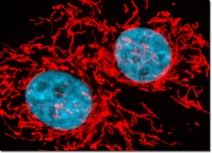
Live tahr ovary cells were treated with MitoTracker Red CMXRos in media containing calf serum for one hour followed by fixation with paraformaldehyde. The fixed cells were then counterstaned with Hoechst 33258 to highlight nuclei in order to examine the spatial relationship between mitochondria and the nucleus in this cell line. Images were recorded in grayscale with a Hamamatsu ORCA-AG camera system coupled to an Olympus BX-51 microscope equipped with bandpass emission fluorescence filter optical blocks provided by Omega Optical. During the processing stage, individual image channels were pseudocolored with RGB values corresponding to each of the fluorophore emission spectral profiles.