Fluorescence Microscopy Digital Image Gallery
Madin-Darby Canine Kidney Epithelial Cells (MDCK Line)
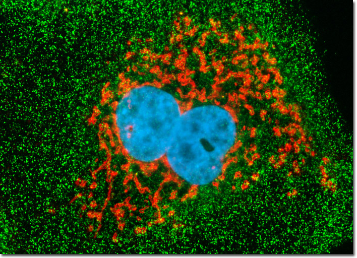
Alexa Fluor dyes were employed to visualize distribution of clathrin and the peroxisomal membrane protein 70 (PMP 70) in the culture of Madin-Darby canine kidney epithelial cells illustrated above. After the immunofluorescence reactions, the cells were counterstained with Hoechst 33342 to reveal the location of the nuclei. Images were recorded in grayscale with a Hamamatsu ORCA-AG camera system coupled to an Olympus BX-51 microscope equipped with bandpass emission fluorescence filter optical blocks provided by Semrock. During the processing stage, individual image channels were pseudocolored with RGB values corresponding to each of the fluorophore emission spectral profiles.