
Image Galleries


Featured Article
 Electron Multiplying Charge-Coupled Devices (EMCCDs)
By incorporating on-chip multiplication gain, the electron multiplying CCD achieves, in an all solid-state sensor, the single-photon detection sensitivity typical of intensified or electron-bombarded CCDs at much lower cost and without compromising the quantum efficiency and resolution characteristics of the conventional CCD structure.
Product Information

Digital Image Gallery
Fluorescence Microscopy Digital Image Gallery
Madin-Darby Canine Kidney Epithelial Cells (MDCK Line)
Derived by S. H. Madin and N. B. Darby from the kidney tissue of an adult female cocker spaniel, the MDCK cell line originated in September 1958. Since that time, the cells have been widely utilized to investigate the processing of beta-amyloid precursor protein, as well as the sorting of its proteolytic products.
The morphology of the MDCK cell line is epithelial, and the cells are positive for keratin as determined by immunoperoxidase staining. Viruses that MDCK cells are susceptible to include vesicular stomatitis (Indiana strain), vaccinia, coxsackievirus B5, reovirus 2 and 3, adenovirus 4 and 5, vesicular exanthema of swine, and infectious canine hepatitis. The cells exhibit resistance to coxsackievirus B3 and B4 as well as poliovirus 2, and are negative for reverse transcriptase.
The MDCK line is commonly used as a general model for epithelial cells, which comprise the type of tissue known as epithelium. Chiefly found covering the internal organs and other surfaces of the body, epithelium is comprised of tightly packed cells that are organized into sheets. These cells secrete an extracellular matrix called the basal lamina at their base, which helps anchor the epithelial tissue to adjacent tissues. Epithelial cells also lack direct access to blood vessels and must, therefore, obtain oxygen and nutrients through diffusion, the same way that they are forced to rid themselves of metabolic waste products. Epithelia function in a variety of mechanisms, including protection, absorption, sensory reception, and secretion. The epithelial cells of the kidneys play a key role in the temporary storage and subsequent secretion of excretory materials.
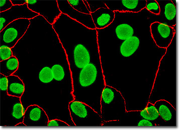
In the digital image presented above, epithelial cell tight junctions and nuclear pore complex proteins were simultaneously imaged in MDCK cells with a cocktail of mouse anti-NPCP and rabbit anti-ZO-3 primary antibodies, followed by goat anti-mouse and anti-rabbit secondary antibodies conjugated to Alexa Fluor 488 and Alexa Fluor 568, respectively. Images were recorded in grayscale with a Hamamatsu ORCA-AG camera system coupled to an Olympus BX-51 microscope equipped with bandpass emission fluorescence filter optical blocks provided by Semrock. During the processing stage, individual image channels were pseudocolored with RGB values corresponding to each of the fluorophore emission spectral profiles.
Additional Fluorescence Images of Madin-Darby Canine Kidney (MDCK) Cells
Staining Actin, Mitochondria, and the Nucleus in MDCK Cells - The adherent culture of Madin-Darby canine kidney cells presented in this section was labeled for the intracellular mitochondrial network and for filamentous actin with MitoTracker Red CMXRos and Alexa Fluor 488 conjugated to phalloidin, respectively. The ultraviolet-absorbing probe DAPI was utilized to counterstain DNA.
Visualizing the Mitochondrial Network in Canine Kidney Cells - A log phase culture of Madin-Darby canine kidney cells was treated with MitoTracker Red CMXRos for one hour, fixed with paraformaldehyde, and counterstained with Hoechst 33258 to highlight the proximal relationship between mitochondria and the nucleus in these epithelial cells. After mounting, the specimen was imaged with a Hamamatsu ORCA-AG digital camera system.
Immunofluorescence Targeting of Tubulin and Peroxisomes in MDCK Cells - Microtubules and peroxisomes were stained using immunofluorescence by treating a fixed and permeabilized culture of MDCK cells with mouse-anti-alpha-tubulin and rabbit anti-PMP 70 (peroxisomal membrane protein) primary antibodies followed by goat anti-mouse antibodies conjugated to Alexa Fluor 568 and Cy5, respectively. Nuclei were counterstained with Hoechst 33342.
Proximity of the Nucleus and Golgi Complex in Monolayer MDCK Cultures - The close proximity between the Golgi complex and nuclei in Madin-Darby canine kidney cells was probed in a double immunofluorescence experiment with mouse anti-NPCP (nuclear pore complex protein) and rabbit anti-giantin (Golgi complex) primary antibodies. The antibody targets were visualized with goat secondary antibodies conjugated to Alexa Fluor 568 and Alexa Fluor 488.
Visualizing the Golgi Complex in Epithelial Cells - The culture of Madin-Darby canine kidney epithelial cells appearing in this section was fixed with paraformaldehyde, permeabilized, and treated with rabbit (anti-giantin) primary antibodies, followed by goat anti-rabbit Fab secondary antibody fragments conjugated to Alexa Fluor 568. Nuclei were visualized by counterstaining the fixed culture with DAPI prior to mounting.
MDCK Cells with Alexa Fluor 488, Alexa Fluor 568, and Hoechst 33342 - Alexa Fluor dyes were employed to visualize distribution of clathrin and the peroxisomal membrane protein 70 (PMP 70) in the culture of Madin-Darby canine kidney epithelial cells illustrated in this section. After the immunofluorescence reactions, the cells were counterstained with Hoechst 33342 to reveal the location of the nuclei.
The Microtubule Network in Epithelial Cells - Microtubules were stained using immunofluorescence by treating a fixed and permeabilized culture of MDCK cells with mouse-anti-alpha-tubulin primary antibodies followed by goat anti-mouse antibodies conjugated to Alexa Fluor 488 (pseudocolored green). Nuclei (pseudocolored cyan) were counterstained with Hoechst 33342.