Fluorescence Microscopy Digital Image Gallery
Madin-Darby Canine Kidney Epithelial Cells (MDCK Line)
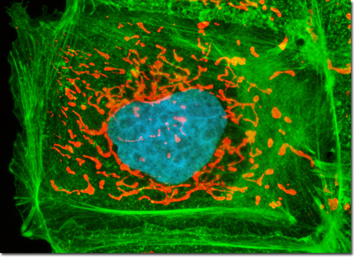
The adherent culture of Madin-Darby canine kidney cells presented in the digital image above was labeled for the intracellular mitochondrial network and for filamentous actin with MitoTracker Red CMXRos and Alexa Fluor 488 conjugated to phalloidin, respectively. The ultraviolet-absorbing probe DAPI was utilized to counterstain DNA. Images were recorded in grayscale with a Hamamatsu ORCA-AG camera system coupled to an Olympus BX-51 microscope equipped with bandpass emission fluorescence filter optical blocks provided by Semrock. During the processing stage, individual image channels were pseudocolored with RGB values corresponding to each of the fluorophore emission spectral profiles.