
Image Galleries


Featured Article
 Electron Multiplying Charge-Coupled Devices (EMCCDs)
By incorporating on-chip multiplication gain, the electron multiplying CCD achieves, in an all solid-state sensor, the single-photon detection sensitivity typical of intensified or electron-bombarded CCDs at much lower cost and without compromising the quantum efficiency and resolution characteristics of the conventional CCD structure.
Product Information

Digital Image Gallery
Fluorescence Microscopy Digital Image Gallery
Indian Muntjac Deer Skin Fibroblast Cells
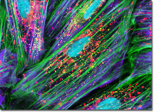
The Indian Muntjac fibroblast cell culture featured in this section was stained with a cocktail of five separate fluorophores to reveal the localization proximity between the nucleus, mitochondria, and peroxisomes, as well as the filamentous actin and vimentin intermediate filament networks. The vimentin network and peroxisomes were immunofluorescently labeled with mouse anti-PMP 70 (a peroxisomal membrane protein) and chicken anti-vimentin primary antibodies followed by secondary Fab fragments conjugated to Alexa Fluor 647 and 750 (pseudocolored yellow and purple, respectively). The filamentous actin network was labeled with Alexa Fluor 488 conjugated to phalloidin (green), while the nuclei and mitochondria were labeled with Hoechst 33342 and MitoTracker Red CMXRos (cyan and red). Images were recorded in grayscale with a Hamamatsu ORCA-AG camera system coupled to an Olympus BX-51 microscope equipped with bandpass emission fluorescence filter optical blocks provided by Semrock. During the processing stage, individual image channels were pseudocolored with RGB values corresponding to each of the fluorophore emission spectral profiles.