Image Galleries
Featured Article
 Electron Multiplying Charge-Coupled Devices (EMCCDs)
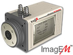
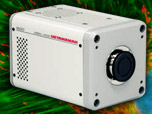
By incorporating on-chip multiplication gain, the electron multiplying CCD achieves, in an all solid-state sensor, the single-photon detection sensitivity typical of intensified or electron-bombarded CCDs at much lower cost and without compromising the quantum efficiency and resolution characteristics of the conventional CCD structure.
Product Information
Digital Video Gallery
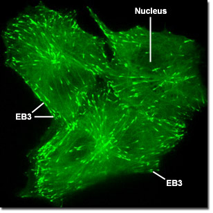
Gray Fox Lung Fibroblast Cells with mEGFP and EB3

Microtubules transport vesicles, organelles, and other molecules throughout the cell. Distribution is regulated by motor proteins that ferry, for instance, secretory vesicles for export from the Golgi apparatus to the plasma membrane. Motor proteins "burn" adenosine triphosphate to fuel the transformation and travel along the microtubules. By altering their three-dimensional conformation, motor proteins move along the microtubule filament by releasing one portion of the filament and gripping another site farther along the tubule. The digital videos in this section illustrate the tracking of microtubule +TIPs (plus end tracking proteins) in Gray fox lung fibroblast cells labeled with a chimera of EB3 fused to mEGFP.
Video 1 - Run Time: 24 Seconds - A trio of fibroblast cells are imaged to reveal fluorescent EB3 emanating from the centrioles. Choose a playback version: Streaming Video (4.7 MB), Progressive Download (4.7 MB), and MPEG Download (45 MB)
Video 2 - Run Time: 17 Seconds - Several fox lung cells display their cytoskeletal networks highlighted by EB3. Choose a playback version: Streaming Video (3.1 MB), Progressive Download (3.1 MB), and MPEG Download (31.1 MB)
Video 3 - Run Time: 40 Seconds - EB3 traversing the cytoplasm of two fox lung fibroblast cells clearly reveals a single origin in each cell. Choose a playback version: Streaming Video (7.4 MB), Progressive Download (7.4 MB), and MPEG Download (70.4 MB)
Video 4 - Run Time: 12 Seconds - A bi-nucleated cell is captured expressing mEGFP-EB3 that outlines the microtubule network. Choose a playback version: Streaming Video (2.8 MB), Progressive Download (2.8 MB), and MPEG Download (27.7 MB)


