Image Galleries
Featured Article
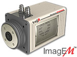
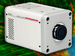
 Electron Multiplying Charge-Coupled Devices (EMCCDs)
By incorporating on-chip multiplication gain, the electron multiplying CCD achieves, in an all solid-state sensor, the single-photon detection sensitivity typical of intensified or electron-bombarded CCDs at much lower cost and without compromising the quantum efficiency and resolution characteristics of the conventional CCD structure.
Product Information
Digital Video Gallery
Epithelial Cell Mitosis
| Exposure: 100 ms | |
| Gain: 3 | |
| Interval: 7 min |
Almost immediately after the metaphase chromosomes are aligned at the metaphase plate, the two halves of each chromosome are pulled apart by the spindle apparatus and migrate to the opposite spindle poles. The kinetochore microtubules shorten as the chromosomes are pulled toward the poles, while the polar microtubules elongate to assist in the separation. Anaphase typically is a rapid process that lasts only a few minutes. When the chromosomes have completely migrated to the spindle poles, the kinetochore microtubules begin to disappear, although the polar microtubules continue to elongate. This is the junction between late anaphase and early telophase, the last stage in chromosome division. In photomicrographs of the process, polar microtubules are in a clearly formed network and the synthesis of a new cell membrane is initiated in the cytoplasm between the two spindle poles. In the digital video presented in this section, pig kidney epithelial cells (LLC-PK1 line) expressing mCherry fluorescent protein fused to histone H2B (red fluorescence) and mEGFP fused to tubulin (green fluorescence) were imaged in 7-minute intervals during mitosis.


