
Image Galleries


Featured Article
 Electron Multiplying Charge-Coupled Devices (EMCCDs)
By incorporating on-chip multiplication gain, the electron multiplying CCD achieves, in an all solid-state sensor, the single-photon detection sensitivity typical of intensified or electron-bombarded CCDs at much lower cost and without compromising the quantum efficiency and resolution characteristics of the conventional CCD structure.
Product Information

Digital Video Gallery
Gray Fox Lung Fibroblast Cells with mKusabira Orange and EB3
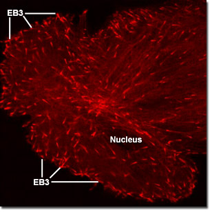
Microtubules are biopolymers that are composed of subunits made from an abundant globular cytoplasmic protein known as tubulin. Each subunit of the microtubule is made of two slightly different but closely related simpler units called alpha-tubulin and beta-tubulin that are bound very tightly together to form heterodimers. The subunits are organized to form 13 parallel protofilaments that point the same direction. This organization gives the structure polarity, with only the alpha-tubulin proteins exposed at one end and only beta-tubulin proteins at the other. The digital videos in this section illustrate the tracking of microtubule +TIPs (plus end tracking proteins) in Gray fox lung fibroblast cells labeled with a chimera of EB3 fused to the fluorescent protein monomeric Kusabira Orange (mKO).
Video 1 - Run Time: 20 Seconds - Two large epithelial cells express mKO fused to EB3 revealing how the chimera migrates to the edges of the cytoplasm. Choose a playback version: Streaming Video (3.7 MB), Progressive Download (3.7 MB), and MPEG Download (36.3 MB)
Video 2 - Run Time: 13 Seconds - A single cell is imaged to reveal fluorescent EB3 radiating through the cytoplasm. Choose a playback version: Streaming Video (2.4 MB), Progressive Download (2.4 MB), and MPEG Download (24.2 MB)
Video 3 - Run Time: 20 Seconds - mKusabira Orange-labeled microtubule ends traverse the cytoplasm in all directions. Choose a playback version: Streaming Video (3.7 MB), Progressive Download (3.7 MB), and MPEG Download (36.3 MB)
Video 4 - Run Time: 20 Seconds - A pair of fox lung cells are captured expressing a fusion mKO and EB3 within the confines of the microtubule network. Choose a playback version: Streaming Video (3.7 MB), Progressive Download (3.7 MB), and MPEG Download (36.3 MB)
Video 5 - Run Time: 20 Seconds - A single fox lung cell reveals its cytoskeletal network through the use of EB3 labeled with monomeric Kusabira Orange fluorescent protein. Choose a playback version: Streaming Video (3.7 MB), Progressive Download (3.7 MB), and MPEG Download (36.3 MB)
Video 6 - Run Time: 10 Seconds - A fibroblast cell is imaged to show fluorescent EB3 traversing through the cytoplasm. Choose a playback version: Streaming Video (1.8 MB), Progressive Download (1.8 MB), and MPEG Download (19 MB)
Video 7 - Run Time: 15 Seconds - mKusabira orange-labeled microtubules move slowly across the cytoplasm. Choose a playback version: Streaming Video (2.8 MB), Progressive Download (2.8 MB), and MPEG Download (27.7 MB)
Video 8 - Run Time: 13 Seconds - mKusabira orange-labeled microtubules emanate from the center of a fox lung cell. Choose a playback version: Streaming Video (2.4 MB), Progressive Download (2.4 MB), and MPEG Download (36.3 MB)
Video 9 - Run Time: 20 Seconds - mKusabira orange-labeled microtubule ends travel in all directions in the cytoplasm of a fox lung cell. Choose a playback version: Streaming Video (3.7 MB), Progressive Download (3.7 MB), and MPEG Download (36.3 MB)