Image Galleries
Featured Article
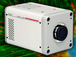
 Electron Multiplying Charge-Coupled Devices (EMCCDs)
By incorporating on-chip multiplication gain, the electron multiplying CCD achieves, in an all solid-state sensor, the single-photon detection sensitivity typical of intensified or electron-bombarded CCDs at much lower cost and without compromising the quantum efficiency and resolution characteristics of the conventional CCD structure.
Product Information
Digital Video Gallery
Cameleon Biosensors in Action
| Exposure: 600 ms | |
| Gain: 3 | |
| Interval: 500 ms |
Perhaps the most widely used biosensor design to screen new or improved FRET pairs involves a protease cleavage assay. The simple motif consists of two fluorescent proteins linked together by a short peptide that contains a consensus protease cleavage site. In general, the sensor exhibits very strong energy transfer that is completely abolished upon cleavage of the linker sequence. Because the technique usually features high dynamic range levels, it can be used to screen new cyan and green FRET donors with yellow, orange, and red acceptors. The largest family of protease biosensors incorporates a cleavage site sensitive to one of the caspase family of proteases, which enables the sensor to be examined during induction of apoptosis. Over the past several years, a large number of novel biosensors using both sensitized fluorescent proteins and FRET pairs have been reported. Despite the continued limitations in dynamic range of FRET sensors using ECFP and EYFP derivatives, this strategy has been widely adopted, probably due to the simplicity of ratiometric measurements and ease of probe construction. New strategies will no doubt emerge using more advanced fluorescent protein combinations that serve to increase the dynamic range and other properties of this highly useful class of probes. In this digital video, a cameleon biosensor composed of cyan and yellow fluorescent proteins sandwiching a calmodulin domain and the m13 domain is being expressed transiently in human cervical carcinoma cells (HeLa line).


