
Image Galleries


Featured Article
 Electron Multiplying Charge-Coupled Devices (EMCCDs)
By incorporating on-chip multiplication gain, the electron multiplying CCD achieves, in an all solid-state sensor, the single-photon detection sensitivity typical of intensified or electron-bombarded CCDs at much lower cost and without compromising the quantum efficiency and resolution characteristics of the conventional CCD structure.
Product Information

Digital Video Gallery
Imaging the Golgi Complex
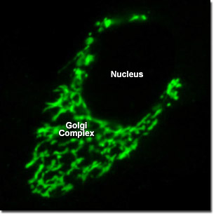
Proteins, carbohydrates, phospholipids, and other molecules formed in the endoplasmic reticulum are transported to the Golgi apparatus to be biochemically modified during their transition from the cis to the trans poles of the complex. Enzymes present in the Golgi lumen modify the carbohydrate (or sugar) portion of glycoproteins by adding or subtracting individual sugar monomers. In addition, the Golgi apparatus manufactures a variety of macromolecules on its own, including a variety of polysaccharides. The digital videos presented in this section examine opossum kidney epithelial cells (OK line) expressing a fusion of mEGFP with a targeting signal to localize the fluorescent protein to the Golgi apparatus.
Video 1 - Run Time: 15 Seconds - Motion in the Golgi sacs is evident surrounding the nucleus in a single opossum kidney cell. Choose a playback version: Streaming Video (2.8 MB), Progressive Download (2.8 MB), and MPEG Download (27.7 MB)
Video 2 - Run Time: 15 Seconds - An extensive Golgi network encapsulates the nucleus of a fibroblast cell and spreads into the cytoplasm. Choose a playback version: Streaming Video (2.8 MB), Progressive Download (2.8 MB), and MPEG Download (27.7 MB)
Video 3 - Run Time: 15 Seconds - A bi-nucleated cell shares Golgi markers. Several rapidly moving particles can be observed in the cytoplasm. Choose a playback version: Streaming Video (2.8 MB), Progressive Download (2.8 MB), and MPEG Download (27.7 MB)
Video 4 - Run Time: 15 Seconds - The Golgi complex completely surrounds an almost spherical nucleus in a single opossum kidney cell. Choose a playback version: Streaming Video (2.8 MB), Progressive Download (2.8 MB), and MPEG Download (27.7 MB)
Video 5 - Run Time: 15 Seconds - Rapid motion is seen in some regions of the Golgi complex in a bi-nucleated opossum kidney fibroblast cell. Choose a playback version: Streaming Video (2.8 MB), Progressive Download (2.8 MB), and MPEG Download (27.7 MB)