Fluorescence Microscopy Digital Image Gallery
Embryonic Rat Thoracic Aorta Cells (A7r5 Line)
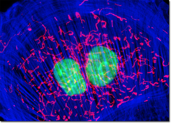
The culture of adherent rat thoracic aorta cells presented above was fluorescently triple-labeled with MitoTracker Red CMXRos, Alexa Fluor 350 conjugated to phalloidin, and SYTOX Green, targeting the mitochondria, filamentous actin network, and nuclei, respectively. In this image, the bright red mitochondrial network is superimposed on a deep blue actin cytoskeletal framework centered around the green nuclei. Images were recorded in grayscale with a Hamamatsu ORCA-AG camera system coupled to an Olympus BX-51 microscope equipped with bandpass emission fluorescence filter optical blocks provided by Semrock. During the processing stage, individual image channels were pseudocolored with RGB values corresponding to each of the fluorophore emission spectral profiles.