Fluorescence Microscopy Digital Image Gallery
Tahr Ovary Epithelial Cells (HJ1.Ov)
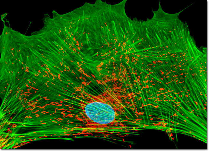
By applying the popular triple-fluorophore combination of MitoTracker Red CMXRos, Alexa Fluor 488 conjugated to phalloidin, and Hoechst 33258 to the culture of normal tahr ovary cells illustrated above, the filamentous actin (green) and mitochondrial (red) networks are revealed, as is the location of the nucleus (blue). Images were recorded in grayscale with a Hamamatsu ORCA-AG camera system coupled to an Olympus BX-51 microscope equipped with bandpass emission fluorescence filter optical blocks provided by Omega Optical. During the processing stage, individual image channels were pseudocolored with RGB values corresponding to each of the fluorophore emission spectral profiles.