Fluorescence Microscopy Digital Image Gallery
Tahr Ovary Epithelial Cells (HJ1.Ov)
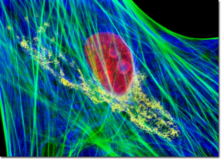
In a double immunofluorescence labeling protocol, adherent tahr ovary cells were fixed, permeabilized, and treated with a cocktail of mouse-anti-vimentin and rabbit anti-giantin primary antibodies, targeting the intermediate filaments and Golgi complex, respectively, followed by goat anti-mouse and anti-rabbit secondary antibodies conjugated to Alexa Fluor 405 (pseudocolored blue) and Alexa Fluor 568 (pseudocolored yellow). The culture was counterstained with Alexa Fluor 488 conjugated to phalloidin (filamentous actin; green) and TO-PRO-3 (nucleus; red) to provide four colors. Images were recorded in grayscale with a Hamamatsu ORCA-AG camera system coupled to an Olympus BX-51 microscope equipped with bandpass emission fluorescence filter optical blocks provided by Omega Optical. During the processing stage, individual image channels were pseudocolored with RGB values corresponding to each of the fluorophore emission spectral profiles.