Fluorescence Microscopy Digital Image Gallery
Tahr Ovary Epithelial Cells (HJ1.Ov)
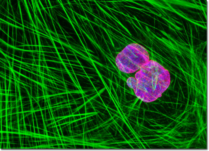
The adherent culture of normal tahr ovary cells presented above was fixed, permeabilized, blocked with 10-percent normal goat serum, and treated with mouse anti-NPCP (nuclear pore complex protein) primary antibodies followed by goat anti-mouse secondary antibodies (IgG) conjugated to Alexa Fluor 568 (red fluorescence). Subsequently, the filamentous actin network and nuclear DNA were labeled with Alexa Fluor 488 conjugated to phalloidin and Hoechst 33258, respectively. Images were recorded in grayscale with a Hamamatsu ORCA-AG camera system coupled to an Olympus BX-51 microscope equipped with bandpass emission fluorescence filter optical blocks provided by Omega Optical. During the processing stage, individual image channels were pseudocolored with RGB values corresponding to each of the fluorophore emission spectral profiles.