Fluorescence Microscopy Digital Image Gallery
Tahr Ovary Epithelial Cells (HJ1.Ov)
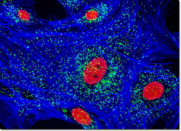
In a double immunofluorescence experiment, fixed and permeabilized adherent HJ1.Ov cells were treated with a cocktail of mouse anti-histones (pan) and rabbit anti-PMP 70 (peroxisomal membrane protein) primary antibodies, followed by a second mixture of goat anti-mouse and anti-rabbit secondary antibodies conjugated to Alexa Fluor 568 and Alexa Fluor 488, respectively (targeting the nucleus and peroxisomes). The filamentous actin cytoskeletal network in the cell culture illustrated above was stained with Alexa Fluor 350 conjugated to phalloidin. Images were recorded in grayscale with a Hamamatsu ORCA-AG camera system coupled to an Olympus BX-51 microscope equipped with bandpass emission fluorescence filter optical blocks provided by Omega Optical. During the processing stage, individual image channels were pseudocolored with RGB values corresponding to each of the fluorophore emission spectral profiles.