Fluorescence Microscopy Digital Image Gallery
Tahr Ovary Epithelial Cells (HJ1.Ov)
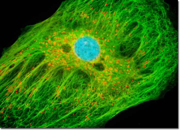
The adherent log phase culture of tahr ovary cells illustrated above was treated for one hour with MitoTracker Red CMXRos in order to label the mitochondrial network, and the fixed cells were then incubated with mouse anti-cytokeratin primary antibodies followed by goat anti-mouse secondary antibodies (IgG) conjugated to Alexa Fluor 488. The nuclei were counterstained with Hoechst 33258. Images were recorded in grayscale with a Hamamatsu ORCA-AG camera system coupled to an Olympus BX-51 microscope equipped with bandpass emission fluorescence filter optical blocks provided by Omega Optical. During the processing stage, individual image channels were pseudocolored with RGB values corresponding to each of the fluorophore emission spectral profiles.