Fluorescence Microscopy Digital Image Gallery
Tahr Ovary Epithelial Cells (HJ1.Ov)
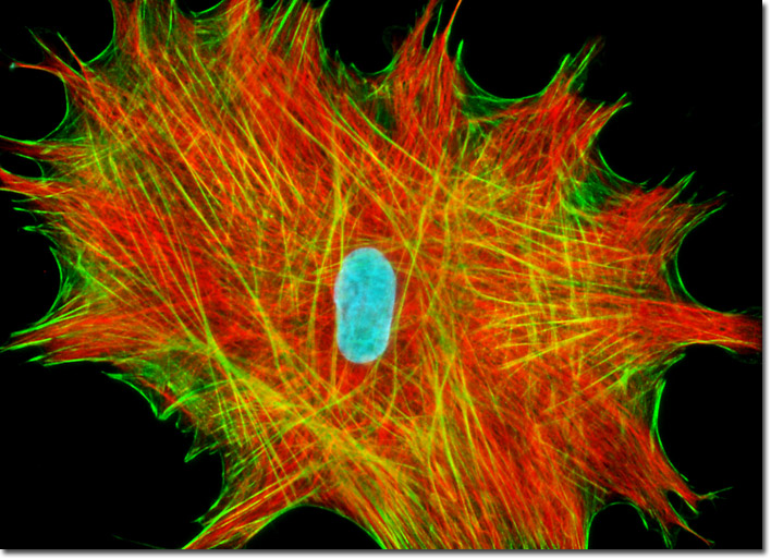
An adherent culture of Tahr Ovary epithelial cells was fixed, permeabilized, and treated with mouse anti-vimentin primary antibodies followed by goat anti-mouse secondary antibody Fab fragments conjugated to Cy3 (pseudocolored red in the digital image presented above). The culture was subsequently counterstained with Alexa Fluor 488 conjugated to phalloidin (to highlight filamentous actin; green) and Hoechst 33342 (nuclei; cyan). Images were recorded in grayscale with a Hamamatsu ORCA-AG camera system coupled to an Olympus BX-51 microscope equipped with bandpass emission fluorescence filter optical blocks provided by Omega Optical. During the processing stage, individual image channels were pseudocolored with RGB values corresponding to each of the fluorophore emission spectral profiles.