Fluorescence Microscopy Digital Image Gallery
Madin-Darby Canine Kidney Epithelial Cells (MDCK Line)
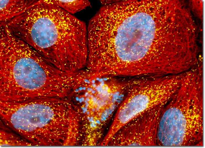
Microtubules and peroxisomes were stained using immunofluorescence by treating a fixed and permeabilized culture of MDCK cells with mouse-anti-alpha-tubulin and rabbit anti-PMP 70 (peroxisomal membrane protein) primary antibodies followed by goat anti-mouse (or rabbit) antibodies conjugated to Alexa Fluor 568 (pseudcolored red) and Cy5 (pseudocolored yellow), respectively. Nuclei were counterstained with Hoechst 33342 (cyan). Note the mitotic cell present in the lower central portion of the window. Images were recorded in grayscale with a Hamamatsu ORCA-AG camera system coupled to an Olympus BX-51 microscope equipped with bandpass emission fluorescence filter optical blocks provided by Semrock. During the processing stage, individual image channels were pseudocolored with RGB values corresponding to each of the fluorophore emission spectral profiles.