Fluorescence Microscopy Digital Image Gallery
Madin-Darby Canine Kidney Epithelial Cells (MDCK Line)
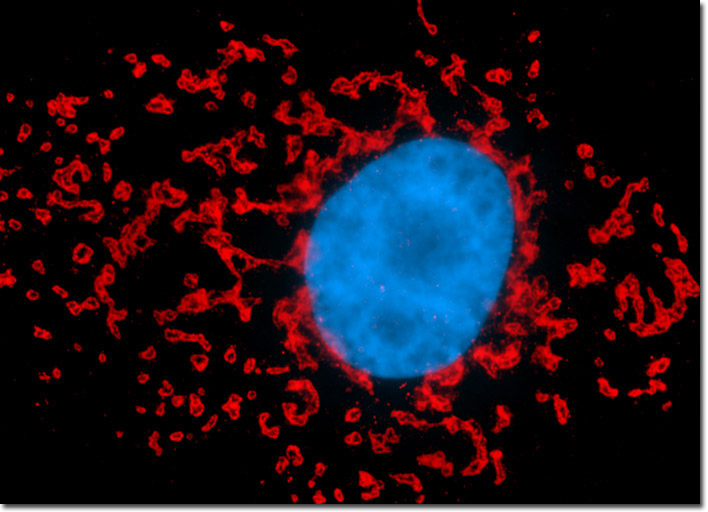
The culture of Madin-Darby canine kidney epithelial cells presented in the image above was fixed with paraformaldehyde, permeabilized, and treated with rabbit (anti-giantin) primary antibodies, followed by goat anti-rabbit Fab secondary antibody fragments conjugated to Alexa Fluor 568. Nuclei were visualized by counterstaining the fixed culture with DAPI prior to mounting. Images were recorded in grayscale with a Hamamatsu ORCA-AG camera system coupled to an Olympus BX-51 microscope equipped with bandpass emission fluorescence filter optical blocks provided by Semrock. During the processing stage, individual image channels were pseudocolored with RGB values corresponding to each of the fluorophore emission spectral profiles.