Fluorescence Microscopy Digital Image Gallery
Transformed African Green Monkey Kidney Fibroblast Cells (COS-7 Line)
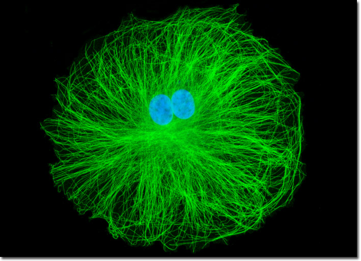
The single cell featured in the digital image above was resident in a COS-7 culture immunofluorescently labeled with primary anti-tubulin mouse monoclonal antibodies followed by goat anti-mouse Fab fragments conjugated to fluorescein isothiocyanate (FITC), which has an absorption maximum of 495 nanometers. The culture was counterstained for DNA in the cell nucleus with the ultraviolet-absorbing probe DAPI. Images were recorded in grayscale with a Hamamatsu ORCA-AG camera system coupled to an Olympus BX-51 microscope equipped with bandpass emission fluorescence filter optical blocks provided by Semrock. During the processing stage, individual image channels were pseudocolored with RGB values corresponding to each of the fluorophore emission spectral profiles.