
Image Galleries


Featured Article
 Electron Multiplying Charge-Coupled Devices (EMCCDs)
By incorporating on-chip multiplication gain, the electron multiplying CCD achieves, in an all solid-state sensor, the single-photon detection sensitivity typical of intensified or electron-bombarded CCDs at much lower cost and without compromising the quantum efficiency and resolution characteristics of the conventional CCD structure.
Product Information

Digital Image Gallery
Fluorescence Microscopy Digital Image Gallery
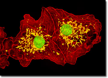
Transformed African Green Monkey Kidney Fibroblast Cells (COS-7 Line)
The COS-7 cell line was derived by Yakov Gluzman in the early 1980s from the previously established CV-1 African green monkey kidney line by transformation of the normal cells with an origin defective mutant of simian virus 40 (SV40) that codes for the wild-type virus T-antigen. The fibroblast line grows adherently to glass and plastic in culture and is generally utilized as a transfection host.
COS-7 cells are susceptible to SV40 (tsA209 strain) at 40 degrees Celsius, and SV40 mutants with deletions in certain regions. Because of this susceptibility, COS-7 cells have been utilized extensively in research, and the cell line is often an excellent choice for transfection experiments with recombinant plasmids. In addition, SV40 itself is a popular subject of study largely due to the fact that the small DNA tumor virus features a relatively simple genetic organization and a genome that can be manipulated with relative ease. Moreover, SV40 has been of exceptional scientific interest due to its possible association with certain types of human cancers, especially mesothelioma.
Enclosed in a spherical capsid, SV40 DNA is 5243 nucleotides in size and exhibits a closed circular superhelical conformation. The capsid that encloses the supercoiled DNA is so small that there is only enough space for the genome to encode a small number of functions. The lifecycle of the virus is controlled by a regulatory portion of the genome, which also encodes three capsid proteins. Space is so limited in the genome that the capsid proteins are encoded with reading frames that overlap, so that the final nucleotide sequence of the gene for one protein also encodes for the initial portion of another protein. In addition to capsid proteins, SV40 DNA encodes the potentially tumor-causing protein T-antigen and a spliced version of the protein called small t-antigen. In primate cells, T-antigen usually controls SV40 DNA replication as well as the packaging of the virus into capsids, but in many other cells the protein binds to p53 and Rb proteins, which are important in the regulation of growth, essentially blocking their normal processes and resulting in the development of tumors.
The culture of transformed African green monkey kidney cells featured in the digital image above was labeled with MitoTracker Red CMXRos (pseudocolored yellow), which selectively binds to targets in the mitochondria. The cells were also stained with Alexa Fluor 568 conjugated to phalloidin and the DNA-binding reagent SYTOX Green, which target the cytoskeletal filamentous actin network and nuclear DNA, respectively. Images were recorded in grayscale with a Hamamatsu ORCA-AG camera system coupled to an Olympus BX-51 microscope equipped with bandpass emission fluorescence filter optical blocks provided by Semrock. During the processing stage, individual image channels were pseudocolored with RGB values corresponding to each of the fluorophore emission spectral profiles.
Additional Fluorescence Images of African Green Monkey Kidney (COS-7) Cells
Visualizing the Cytoskeleton in African Green Monkey Kidney Cells - The adherent COS-7 culture featured in this section was immunofluorescently labeled with primary anti-bovine alpha-tubulin mouse monoclonal antibodies followed by goat anti-mouse Fab fragments conjugated to fluorescein. The culture was counterstained with phalloidin conjugated to Alexa Fluor 568 and DAPI.
COS-7 Cells with MitoTracker Red CMXRos, Alexa Fluor 488, and Hoechst 33258 - A culture of COS-7 fibroblasts was labeled for mitochondria with MitoTracker Red CMXRos, and for the cytoskeletal filamentous actin network with Alexa Fluor 488 conjugated to phalloidin, a cyclic peptide derived from the toxic death cap fungus (Amanita phalloides). In addition, the fibroblasts were counterstained for DNA in the cell nucleus with Hoechst 33258.
Probing the Golgi and Nucleoli in COS-7 Cells - In a double immmunofluorescence experiment, fixed and permeabilized COS-7 cells were treated with a cocktail of mouse anti-fibrillarin and goat anti-giantin primary antibodies, targeting the nucleoli and Golgi complex, respectively. The organelles were visualized with secondary antibody Fab fragments conjugated to Alexa Fluor 488 and 568.
African Green Monkey Kidney Fibroblasts with FITC and DAPI - The single cell featured in this section was resident in a COS-7 culture immunofluorescently labeled with primary anti-tubulin mouse monoclonal antibodies followed by goat anti-mouse Fab fragments conjugated to fluorescein isothiocyanate (FITC), which has an absorption maximum of 495 nanometers. The culture was counterstained for DNA in the cell nucleus with the ultraviolet-absorbing probe DAPI.
Intracellular Mitochondria in African Green Monkey Kidney Cells - In the digital image featured in this section, the mitochondria present in a culture of COS-7 kidney fibroblasts are easily observable due to staining with MitoTracker Deep Red 633. The filamentous actin and cell nuclei present in the culture, which were respectively labeled with Alexa Fluor 488 conjugated to phalloidin and SYTOX Orange, are also readily apparent.
Mapping Actin and the Nucleus in COS-7 Cells - A log-phase, adherent culture of COS-7 cells was fixed, permeabilized, blocked, and treated with phalloidin conjugated to Alexa Fluro 568, highlighting numerous details of the actin cytoskeletal network. After washing, the cells were counterstained with the DNA-binding dye Hoechst 33258 to reveal nuclear proximity to various cytoskeletal elements.
COS-7 Kidney Cells with Cy3, Alexa Fluor 488, and DAPI - An adherent culture of COS-7 African green monkey kidney fibroblast cells was immunofluorescently labeled with primary anti-tubulin mouse monoclonal antibodies followed by goat anti-mouse Fab fragments conjugated to Cy2, targeting microtubules. The cells were simultaneously stained for the cytoskeletal filamentous actin network with Alexa Fluor 568 conjugated to phalloidin, and for DNA with DAPI.