Fluorescence Microscopy Digital Image Gallery
Transformed African Green Monkey Kidney Fibroblast Cells (COS-7 Line)
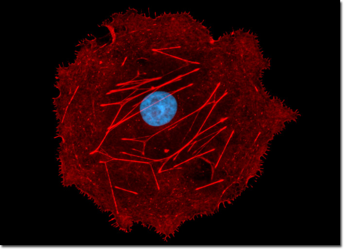
A log-phase, adherent culture of COS-7 cells was fixed, permeabilized, blocked, and treated with phalloidin conjugated to Alexa Fluro 568, highlighting numerous details of the actin cytoskeletal network (pseudocolored red). After washing, the cells were counterstained with the DNA-binding dye Hoechst 33258 (pseudocolored cyan) to reveal nuclear proximity to various cytoskeletal elements. Images were recorded in grayscale with a Hamamatsu ORCA-AG camera system coupled to an Olympus BX-51 microscope equipped with bandpass emission fluorescence filter optical blocks provided by Semrock. During the processing stage, individual image channels were pseudocolored with RGB values corresponding to each of the fluorophore emission spectral profiles.