Fluorescence Microscopy Digital Image Gallery
Indian Muntjac Deer Skin Fibroblast Cells
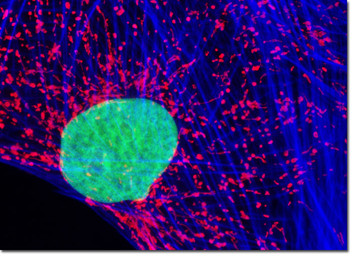
The culture of Indian Muntjac deer skin fibroblast cells presented in the digital image above was labeled for the cytoskeletal filamentous actin network with Alexa Fluor 350 conjugated to phalloidin, and for the cell nucleus with SYTOX Green. Additionally, cellular mitochondria were stained with MitoTracker Red CMXRos, a complex aminated xanthene derivative. Images were recorded in grayscale with a Hamamatsu ORCA-AG camera system coupled to an Olympus BX-51 microscope equipped with bandpass emission fluorescence filter optical blocks provided by Semrock. During the processing stage, individual image channels were pseudocolored with RGB values corresponding to each of the fluorophore emission spectral profiles.