
Image Galleries


Featured Article
 Electron Multiplying Charge-Coupled Devices (EMCCDs)
By incorporating on-chip multiplication gain, the electron multiplying CCD achieves, in an all solid-state sensor, the single-photon detection sensitivity typical of intensified or electron-bombarded CCDs at much lower cost and without compromising the quantum efficiency and resolution characteristics of the conventional CCD structure.
Product Information

Digital Image Gallery
Fluorescence Microscopy Digital Image Gallery
Indian Muntjac Deer Skin Fibroblast Cells
A fibroblast cell line established from a skin biopsy of an adult male, the Indian Muntjac deer epidermis line is commonly used in laboratories around the world, especially for chromosome studies. Members of the family Cervidae, Muntjacs are barking deer that emit their characteristic sound when they feel threatened or alarmed.
The normal (non-transformed) Indian Muntjac cell line is susceptible to the herpes simplex virus, vaccinia virus, and vesicular stomatitis virus (Indiana strain), but is resistant to poliovirus 1. Recent tests have demonstrated that the cells produce both detectable bovine viral diarrhea virus (BVDV) antigens and infectious BVDV virions. Muntjac cells are negative for reverse transcriptase, indicating the lack of integral retrovirus genomes.
Cell lines derived from the Indian Muntjac have been of significant scientific interest primarily because the animal possesses the fewest number of diploid chromosomes of all mammals, with only six chromosomes in the female and seven in the male. Such a small number of chromosomes makes Indian Muntjac cells an ideal candidate for mitosis research. Moreover, in recent years, Indian Muntjac cells have gained a reputation for their usefulness as a model to study telomere biology. Telomeres, the regions of DNA that occur at the end of chromosomes, are at the center of many modern studies involving the aging of organisms and cell senescence. It is generally believed that the shortening of telomeres, which occurs during the division of most cells, is responsible for age-related cellular malfunction and death. Thus, telomeres are also of interest in regard to cancer, since tumor cells are often able to proliferate unhindered by the passage of time due to various mechanisms that avoid the shortening of chromosomal telomeres.
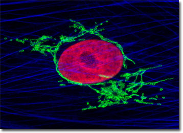
The culture of Muntjac cells illustrated above was triple-labeled using double immunofluorescence and a phallotoxin. Nuclei were visualized with mouse anti-histones (core) primary antibodies, while the Golgi complex was stained with rabbit anti-giantin antibodies. Secondary antibodies were goat anti-mouse and anti-rabbit conjugated to Texas Red and Oregon Green 488, respectively to produce red nuclei and green Golgi cisternae. The filamentous actin network was counterstained with Alexa Fluor 350 conjugated to phalloidin. Images were recorded in grayscale with a Hamamatsu ORCA-AG camera system coupled to an Olympus BX-51 microscope equipped with bandpass emission fluorescence filter optical blocks provided by Semrock. During the processing stage, individual image channels were pseudocolored with RGB values corresponding to each of the fluorophore emission spectral profiles.
Additional Fluorescence Images of Indian Muntjac Deer Skin Fibroblast Cells
Mitochondria, Actin, and Nuclear Localization in Indian Muntjac Cells - The adherent monolayer culture of Indian Muntjac cells illustrated above was immunofluorescently labeled with primary mouse anti-oxphos complex V inhibitor protein antibodies, followed by goat anti-mouse Fab fragments conjugated to Cy2. The culture was subsequently stained with Alexa Fluor 568 conjugated to phalloidin to reveal details of the filamentous actin network, and DAPI for DNA in the nucleus.
Histone and Peroxisome Distribution in Indian Muntjac Cell Cultures - In a double immunofluorescence experiment, the adherent monolayer culture of Indian Muntjac cells presented above was fixed, permeabilized, blocked with 10 percent normal goat serum, and treated with a cocktail of mouse anti-histones (pan) and rabbit anti-PMP 70 (peroxisomal membrane protein) primary antibodies, followed by goat anti-mouse and anti-rabbit secondary antibodies (IgG) conjugated to Texas Red and Oregon Green 488, respectively.
Indian Muntjac Fibroblast Cell Cultures with Cy2, Alexa Fluor 568, and DAPI - The digital image presented in this section features a culture of Indian Muntjac fibroblast cells that was immunofluorescently labeled with primary anti-tubulin mouse monoclonal antibodies followed by goat anti-mouse Fab fragments conjugated to the cyanine dye, Cy2. In addition, the culture was counterstained for DNA with DAPI, and for the cytoskeletal filamentous actin network with Alexa Fluor 568 conjugated to phalloidin.
Probing the Nucleus and Golgi Complex with Indian Muntjac Cells - The Indian Muntjac cells presented in this section were resident in an adherent culture stained for DNA with the bis-benzimidazole dye Hoechst 33258 (blue fluorescence). In addition, the culture was immunofluorescently labeled with Alexa Fluor 488 (green fluorescence) and Alexa Fluor 568 (pseudocolored red) conjugated to goat secondary antibodies that target mouse anti-fibrillarin (nucleoli) and rabbit anti-giantin (targeting the Golgi complex) primary antibodies, respectively.
Indian Muntjac Cells Stained for Actin, Mitochondria, and the Nucleus - The culture of Indian Muntjac deer skin fibroblast cells in this section was labeled for the cytoskeletal filamentous actin network with Alexa Fluor 350 conjugated to phalloidin, and for the cell nucleus with SYTOX Green. Additionally, cellular mitochondria were stained with MitoTracker Red CMXRos, a complex aminated xanthene derivative.
Examining Golgi and Microtubule Elements in Fibroblast Cells - The expansive cellular microtubule network present in a culture of Indian Muntjac fibroblasts was immunofluorescently labeled with primary anti-tubulin mouse monoclonal antibodies and rabbit anti-giantin polyclonial antibodies followed by goat anti-mouse Fab fragments conjugated to Rhodamine Red-X and Alexa Fluor 647, respectively.
Four-Color Fluorescence Labeling of Indian Muntjac Cells - Examination of adherent cells with four-color fluorescence requires fluorophores having emission profiles that span the visible wavelength region. The culture featured in this section was immunofluorescently labeled with Alexa Fluor 647, Oregon Green 488, and Texas Red. Subsequently, the cells were stained with Alexa Fluor 530 conjugated to phalloidin.
Muntjac Fibroblasts with Alexa Fluor 488, MitoTracker Red CMXRos, and DAPI - Indian Muntjac deer skin fibroblast cells were labeled with Alexa Fluor 488 conjugated to phalloidin to target the cytoskeletal filamentous actin network, and for MitoTracker Red CMXRos to visualize the extensive intracellular tubular mitochondria. The cells were subsequently counterstained with DAPI, which targets DNA in the nucleus.
Examining the Relationship Between Microtubules and Peroxisomes - In a double immunofluorescence experiment, an adherent monolayer culture of Indian Muntjac cells was fixed, permeabilized, blocked with 10 percent normal goat serum, and treated with a cocktail of mouse anti-tubulin and rabbit anti-PMP 70 (peroxisomal membrane protein) primary antibodies, followed by goat anti-mouse and anti-rabbit secondary antibodies (IgG) conjugated to Texas Red and Oregon Green 488, respectively.
Labeling Intermediate Filaments and Actin in Muntjac Cells - In order to label the intermediate filaments in the log phase adherent Indian Muntjac culture presented in this section, the fixed and permeabilized cells were blocked and treated with mouse anti-vimentin (porcine eye lens) primary antibodies followed by goat anti-mouse secondary antibodies (IgG) conjugated to Texas Red-X.
Three-Color Cytoskeletal Fluorescence with Indian Muntjac Cells - The digital image presented in this section features a resident cell from a culture of Indian Muntjac fibroblasts that was labeled for DNA with DAPI, and for the cytoskeletal filamentous actin network with Alexa Fluor 568 conjugated to phalloidin, as well as alpha-tubulin in the microtubulin cytoskeleton labeled with Alexa Fluor 488.
Five-Color Fluorescence in Mammalian Cell Cultures - The Indian Muntjac fibroblast cell culture featured in this section was stained with a cocktail of five separate fluorophores to reveal the localization proximity between the nucleus, mitochondria, and peroxisomes, as well as the filamentous actin and vimentin intermediate filament networks. Images were recorded with a Hamamatsu ORCA-AG camera system.
The Golgi Complex in Indian Muntjac Cells - A culture of Indian Muntjac fibroblasts was fixed, permeabilized, and blocked with 10-percent normal goat serum in phosphate-buffered saline prior to immunofluorescent labeling with primary antibodies to giantin, a protein resident in the Golgi complex of mammalian cells. The culture was subsequently stained with secondary antibody fragments (heavy and light chain) conjugated to Cy2. In addition, the culture was labeled for mitochondria with MitoTracker Red CMXRos, and for DNA with Hoechst 33342.
Microtubules, Peroxisomes, and Nuclear Staining in Fibroblast Cells - The culture of Indian Muntjac cells featured in this section were labeled in a dual immunofluorescence experiment with mouse anti-tubulin and rabbit anti-PMP70 primary antibodies followed by secondary antibodies conjugated to Alexa Fluor 568 and 488 respectively. Nuclei were counterstained with Hoechst 33342.