Fluorescence Microscopy Digital Image Gallery
Indian Muntjac Deer Skin Fibroblast Cells
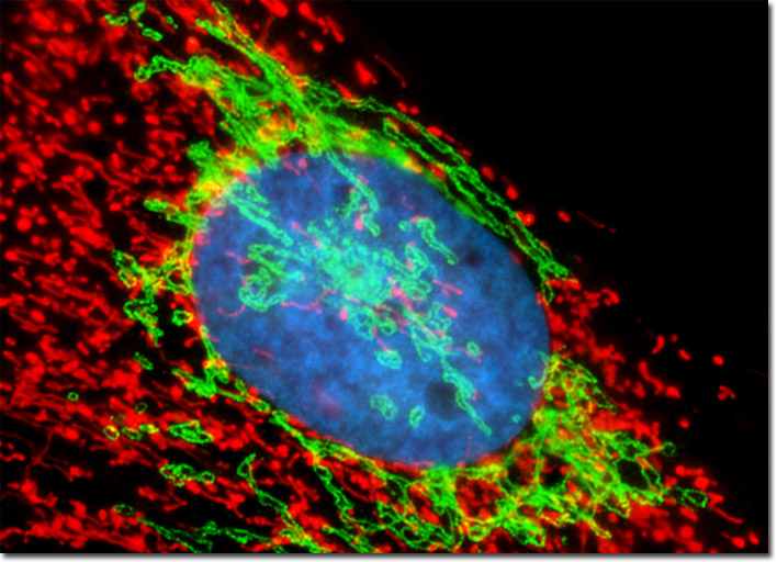
The culture of Indian Muntjac fibroblasts that appears in the digital image above was fixed, permeabilized, and blocked with 10-percent normal goat serum in phosphate-buffered saline prior to immunofluorescent labeling with primary antibodies to giantin, a protein resident in the Golgi complex of mammalian cells. The culture was subsequently stained with secondary antibody fragments (heavy and light chain) conjugated to Cy2. In addition, the culture was labeled for mitochondria with MitoTracker Red CMXRos, and for DNA with Hoechst 33342. Images were recorded in grayscale with a Hamamatsu ORCA-AG camera system coupled to an Olympus BX-51 microscope equipped with bandpass emission fluorescence filter optical blocks provided by Semrock. During the processing stage, individual image channels were pseudocolored with RGB values corresponding to each of the fluorophore emission spectral profiles.