Fluorescence Microscopy Digital Image Gallery
Indian Muntjac Deer Skin Fibroblast Cells
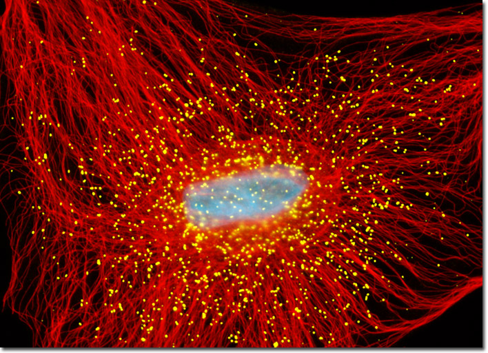
The culture of Indian Muntjac cells featured in this section were labeled in a dual immunofluorescence experiment with mouse anti-tubulin and rabbit anti-PMP70 primary antibodies followed by secondary antibodies conjugated to Alexa Fluor 568 (red) and 488 (yellow) respectively. Nuclei were counterstained with Hoechst 33342 (cyan). Images were recorded in grayscale with a Hamamatsu ORCA-AG camera system coupled to an Olympus BX-51 microscope equipped with bandpass emission fluorescence filter optical blocks provided by Semrock. During the processing stage, individual image channels were pseudocolored with RGB values corresponding to each of the fluorophore emission spectral profiles.