Fluorescence Microscopy Digital Image Gallery
Indian Muntjac Deer Skin Fibroblast Cells
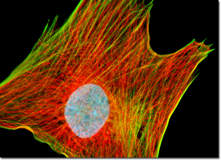
The digital image presented above features a cell culture of Indian Muntjac fibroblasts that was immunofluorescently labeled with primary anti-tubulin mouse monoclonal antibodies followed by goat anti-mouse Fab fragments conjugated to the cyanine dye, Cy2. In addition, the culture was counterstained for DNA with DAPI, and for the cytoskeletal filamentous actin network with Alexa Fluor 568 conjugated to phalloidin. Images were recorded in grayscale with a Hamamatsu ORCA-AG camera system coupled to an Olympus BX-51 microscope equipped with bandpass emission fluorescence filter optical blocks provided by Semrock. During the processing stage, individual image channels were pseudocolored with RGB values corresponding to each of the fluorophore emission spectral profiles.