Fluorescence Microscopy Digital Image Gallery
Indian Muntjac Deer Skin Fibroblast Cells
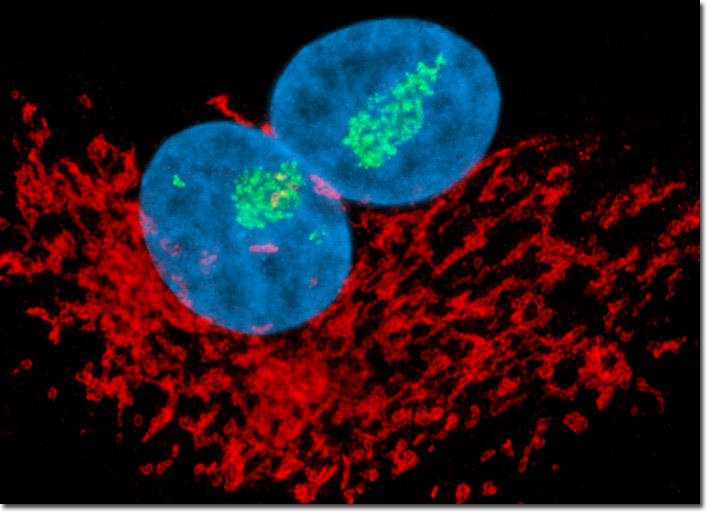
The Indian Muntjac cells presented above were resident in an adherent culture stained for DNA with the bis-benzimidazole dye Hoechst 33258 (blue fluorescence). In addition, the culture was immunofluorescently labeled with Alexa Fluor 488 (green fluorescence) and Alexa Fluor 568 (pseudocolored red) conjugated to goat secondary antibodies that target mouse anti-fibrillarin (nucleoli) and rabbit anti-giantin (targeting the Golgi complex) primary antibodies, respectively. Images were recorded in grayscale with a Hamamatsu ORCA-AG camera system coupled to an Olympus BX-51 microscope equipped with bandpass emission fluorescence filter optical blocks provided by Semrock. During the processing stage, individual image channels were pseudocolored with RGB values corresponding to each of the fluorophore emission spectral profiles.