Fluorescence Microscopy Digital Image Gallery
Indian Muntjac Deer Skin Fibroblast Cells
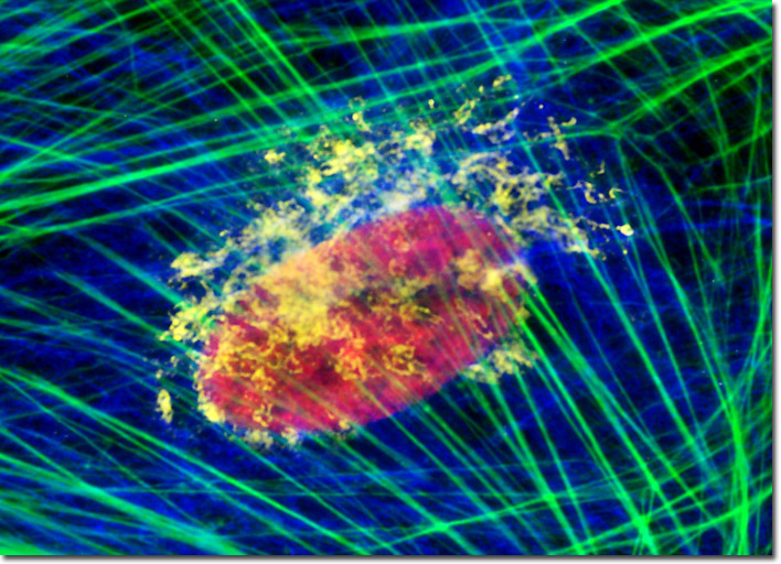
The culture of Muntjac cells illustrated above was quadruple-labeled using triple immunofluorescence and a phallotoxin. Nuclei were visualized with mouse anti-histones (core) primary antibodies, while the Golgi complex was stained with rabbit anti-giantin antibodies. Secondary antibodies were goat anti-mouse and anti-rabbit conjugated to Texas Red and Oregon Green 488, respectively to produce red nuclei and yellow Golgi cisternae. Intermediate filaments were targeted with chicken anti-vimentin primary antibodies followed by goat anti-chicken secondaries conjugated to Alexa Fluor 647 (pseudocolored blue). The filamentous actin network was counterstained with Alexa Fluor 350 conjugated to phalloidin (pseudocolored green). Images were recorded in grayscale with a Hamamatsu ORCA-AG camera system coupled to an Olympus BX-51 microscope equipped with bandpass emission fluorescence filter optical blocks provided by Semrock. During the processing stage, individual image channels were pseudocolored with RGB values corresponding to each of the fluorophore emission spectral profiles.