
Image Galleries


Featured Article
 Electron Multiplying Charge-Coupled Devices (EMCCDs)
By incorporating on-chip multiplication gain, the electron multiplying CCD achieves, in an all solid-state sensor, the single-photon detection sensitivity typical of intensified or electron-bombarded CCDs at much lower cost and without compromising the quantum efficiency and resolution characteristics of the conventional CCD structure.
Product Information

Digital Video Gallery
Human Carcinoma (HeLa) Cells with mEGFP and EB3
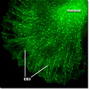
A variety of microtubule binding proteins, including a family of end-binding proteins (known as EBs), have been demonstrated to associate specifically with the ends of growing microtubules in a variety of cell types. These proteins are believed to regulate microtubule dynamics and the binding of microtubules to organelles, membrane components, and other protein complexes. The digital videos below explore the dynamics of microtubule +TIPs (plus end tracking proteins) in human cervical carcinoma (HeLa line) epithelial cells labeled with a chimera of EB3 fused to mEGFP.
Video 1 - Run Time: 19 Seconds - A single cell is imaged to reveal fluorescent EB3 emanating from the centrioles. Choose a playback version: Streaming Video (3.7 MB), Progressive Download (3.7 MB), and MPEG Download (36.1 MB)
Video 2 - Run Time: 10 Seconds - In a cell that is over expressing EB3, the labeled protein highlights microtubules while traversing through the cytoplasm. Choose a playback version: Streaming Video (1.9 MB), Progressive Download (1.9 MB), and MPEG Download (19.1 MB)
Video 3 - Run Time: 39 Seconds - Two cells that appear to have just divided express mEGFP-labeled EB3 from seemingly mirrored centrioles. Choose a playback version: Streaming Video (7.5 MB), Progressive Download (7.5 MB), and MPEG Download (70.8 MB)
Video 4 - Run Time: 25 Seconds - A bi-nucleated cell is captured expressing mEGFP-EB3 that outlines the microtubule network. Choose a playback version: Streaming Video (4.7 MB), Progressive Download (4.7 MB), and MPEG Download (41.6 MB)
Video 5 - Run Time: 28 Seconds - Several HeLa cells display their cytoskeletal networks highlighted by EB3. Choose a playback version: Streaming Video (5.4 MB), Progressive Download (5.4 MB), and MPEG Download (51.1 MB)
Video 6 - Run Time: 35 Seconds - A large epithelial cell extruding numerous pseudopodia illustrates how EB3 migrates to the edges of the cytoplasm. Choose a playback version: Streaming Video (6.6 MB), Progressive Download (6.6 MB), and MPEG Download (62.4 MB)
Video 7 - Run Time: 15 Seconds - EB3-labeled microtubule ends traverse the cytoplasm like fireflies on a summer evening. Choose a playback version: Streaming Video (2.8 MB), Progressive Download (2.8 MB), and MPEG Download (27.8 MB)