Image Galleries
Featured Article
 Electron Multiplying Charge-Coupled Devices (EMCCDs)
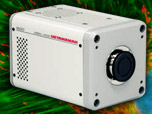
By incorporating on-chip multiplication gain, the electron multiplying CCD achieves, in an all solid-state sensor, the single-photon detection sensitivity typical of intensified or electron-bombarded CCDs at much lower cost and without compromising the quantum efficiency and resolution characteristics of the conventional CCD structure.
Product Information
Digital Video Gallery
Digital Videos of Cells in Culture
The application of fluorescent proteins as probes of intracellular activity has revolutionized the field of cell biology. The digital videos presented in this gallery investigate a wide variety of these ubiquitous probes in fusions with subcellular localization peptides and proteins. A number of epithelial and fibroblast cell lines expressing fluorescent protein fusions are imaged with a Hamamatsu ImageEM electron multiplying camera system coupled to an Olympus DSU spinning disk microscope. Videos are presented in a streaming format, but they can also be downloaded as MPEG files or in a buffered progressive download format for repeated viewing.
Dynamics of Tubulin and Associated Proteins
HeLa cells with EGFP fused to the microtubule-binding protein EB3 - A variety of microtubule binding proteins, including a family of end-binding proteins (known as EBs), have been demonstrated to associate specifically with the ends of growing microtubules in a variety of cell types. These proteins are believed to regulate microtubule dynamics and the binding of microtubules to organelles, membrane components, and other protein complexes.
Fox lung cells with EGFP fused to the microtubule-binding protein EB3 - Microtubules transport vesicles, organelles, and other molecules throughout the cell. Distribution is regulated by motor proteins that ferry, for instance, secretory vesicles for export from the Golgi apparatus to the plasma membrane. Motor proteins "burn" adenosine triphosphate to fuel the transformation and travel along the microtubules. By altering their three-dimensional conformation, motor proteins move along the microtubule filament by releasing one portion of the filament and gripping another site farther along the tubule.
Fox lung cells with mKusabira Orange fused to the microtubule-binding protein EB3 - Microtubules are biopolymers that are composed of subunits made from an abundant globular cytoplasmic protein known as tubulin. Each subunit of the microtubule is made of two slightly different but closely related simpler units called alpha-tubulin and beta-tubulin that are bound very tightly together to form heterodimers. The subunits are organized to form 13 parallel protofilaments that point the same direction. This organization gives the structure polarity, with only the alpha-tubulin proteins exposed at one end and only beta-tubulin proteins at the other.
Fox lung cells with mEGFP fused to tubulin - The ends of microtubules are always in dynamic formation: addition or removal of tubulin proteins alters the length of the biopolymer. The rate of depolymerization is different for each end according to its polarity, and the side that grows the fastest is considered the positive end while the other, more stable terminus, is negative. The dynamic, fast-growing portion of the microtubule is composed of beta-tubulin projected out towards the membrane of the cell. The alpha-tubulins stabilize their structure toward the nucleus at the centriole located in the centrosome of the cell. The digital videos presented in this section explore Gray fox lung fibroblast cells (FoLu line) expressing a fusion construct of mEGFP and human alpha-tubulin to highlight microtubules (green fluorescence) in order to visualize dynamic processes.
Observing Chromosomes and the Spindle in Mitosis
Pig kidney cells with EGFP-Tubulin and mCherry-H2B - The digital video sequences presented in this section feature epithelial cells undergoing mitosis as visualized through the highly dynamic interactions between fluorescently labeled red histones and green tubulin. Cells enter mitosis through prophase and proceed through division to create a pair of daughter cells. The labeled histones enable visualization of the condensed chromatin as it progresses through various phases, while the tubulin can be seen forming the mitotic spindle as well as the midbodies at telophase.
Pig kidney cells with mEmerald-EB3 and mCherry-H2B - The fluorescent protein known as mEmerald is a high-performance version of EGFP that is significantly brighter and also matures faster. When coupled to the microtubule end-binding protein EB3, traces of the fluorescently labeled positive microtubule ends can be observed traversing the cytoplasm and forming the spindle during mitosis. Compare these digital videos with those of EGFP-tubulin presented in the section above.
Mitochondrial Dynamics in Live Cells
Fox lung cells with mEGFP labeled mitochondria - The number of mitochondria present in a cell depends upon the metabolic requirements of that cell, and may range from a single large mitochondrion to thousands of the organelles. In the digital videos presented in this section, normal Gray fox lung fibroblast cells (FoLu line) are expressing monomeric green fluorescent protein (mEGFP) fused to the mitochondrial targeting signal from subunit VIII of human cytochrome C oxidase.
Pig kidney cells expressing TurboRFP fused to a mitochondrial targeting peptide - Although TurboRFP is a dimeric orange fluorescent protein, it can still be effectively localized to the mitochondria when fused to the appropriate targeting signal. Apparently, the dimeric physical character of this extremely bright and photostable fluorescent protein doesn't affect non-specific targeting. However TurboRFP is not properly localized in fusions to proteins that form biopolymers, such as tubulin, connexins, histones, and actin.
Opossum kidney cells with Katushka fluorescent protein mitochondrial expression - Katushka is a derivative of TurboRFP and retains the dimeric character of its parent. When fused to the mitochondrial targeting signal from subunit VIII of human cytochrome C oxidase, however, Katushka fluorescent protein is able to localize to the mitochondria in human and animal cell lines.
Time-Lapse Imaging of the Endoplasmic Reticulum
Human osteosarcoma cells with EYFP expressed in the endoplasmic reticulum - The endoplasmic reticulum is a network of flattened sacs and branching tubules that extends throughout the cytoplasm in plant and animal cells. The digital videos presented in this section examine human osteosarcoma cells (U2OS line) expressing enhanced yellow fluorescent protein (EYFP) targeted to the endoplasmic reticulum with calreticulin and KDEL signal peptides.
Pig kidney cells expressing mEmerald fluorescent protein in the endoplasmic reticulum - The endoplasmic reticulum (ER) is a vast network of flattened sacs and branching tubules that extends throughout the cytoplasm in plant and animal cells. These sacs and tubules are all interconnected by a single continuous membrane so that the organelle has only one large, highly convoluted and complexly arranged lumen (internal space). The digital videos in this section examine the dynamic action of endoplasmic reticulum membranes in cultured pig kidney cells.
The Golgi Complex
Opossum kidney cells with mEGFP expressed in the Golgi complex - Proteins, carbohydrates, phospholipids, and other molecules formed in the endoplasmic reticulum are transported to the Golgi apparatus to be biochemically modified during their transition from the cis to the trans poles of the complex. Enzymes present in the Golgi lumen modify the carbohydrate (or sugar) portion of glycoproteins by adding or subtracting individual sugar monomers. In addition, the Golgi apparatus manufactures a variety of macromolecules on its own, including a variety of polysaccharides.
Imaging Live Cells with Double Labels
Owl monkey kidney cells labeled with mEGFP and DsRed fluorescent proteins - The digital videos in this section examine owl monkey kidney epithelial cells (OMK line) that were transfected with a mixture of mEGFP fused to a mitochondrial targeting signal (green fluorescence) and DsRed fluorescent protein fused to an endoplasmic reticulum targeting signal (red fluorescence). The dual labeling enables the observation of the complex interplay between mitochondria and the endoplasmic reticulum. Mitochondria are similar to plant chloroplasts in that both organelles are able to produce energy and metabolites that are required by the host cell. The endoplasmic reticulum is centrally involved in cellular protein processing.
Human bone cancer cells expressing DsRed and mEGFP fluorescent proteins - First discovered through the use of a light microscope in the 1800s, mitochondria are found in almost all eukaryotes: animals, protists, and even fungi. The scientists who first observed these organelles, because of the way they appeared under the microscope, termed them mitochondria after the Greek words for "granule" and "thread". This break-through led to our current understanding of the functioning of the mitochondrion. In the digital videos presented in this section, a human osteosarcoma cell (U2OS line) is co-expressing DsRed fluorescent protein fused to an endoplasmic reticulum targeting signal and mEGFP fused to a mitochondrial targeting signal.
Fox lung cells with labeled mitochondria and EB3 - The digital videos presented in this section illustrate the interplay between mitochondria (labeled with DsRed fluorescent protein) and the microtubules by examining the tracking of microtubule +TIPs (plus end tracking proteins) labeled with EB3 fused to the popular tracking probe, mEGFP. The length of microtubules in the cell varies between 200 nanometers and 25 micrometers, depending upon the task of a particular microtubule and the state of the cell's life cycle. Microtubules are believed to act as a transport mechanism for mitochondria. EB3 is a protein that binds to the plus-ends of microtubules and can be visualized tracking through the cytoplasm.
Fox lung cells with fluorescent proteins in the mitochondria and Golgi complex - Due to its relatively large size, the Golgi apparatus was one of the first organelles ever observed. In 1897, an Italian physician named Camillo Golgi, who was investigating the nervous system by using a new staining technique he developed (and which is still sometimes used today; known as Golgi staining or Golgi impregnation), observed in a sample under his light microscope, a cellular structure that he termed the "internal reticular apparatus." The digital videos presented in this section illustrate the interplay between mitochondria (labeled with DsRed fluorescent protein) and the Golgi complex (labeled with mEGFP) in Gray fox lung (FoLu line) fibroblast cells.
Annexin Translocation in HeLa Cells
Translocation of annexin (A4) to the membrane in HeLa cells - The annexin family is structurally distinct from other calcium-binding proteins, and each member consists of an N-terminal domain and a typical core domain. The core domain binds calcium and phospholipids and is comprised of four annexin repeats, each being approximately 70 residues in size. This basic core is conserved among all members of the annexin family. Through mediation of intracellular calcium signals, annexins have been demonstrated to play a role in a variety of cellular processes. The digital videos in this section illustrate the effects of calcium induction on mEGFP was fused to annexin A4 and transfected into human cervical carcinoma epithelial cells (HeLa line).
Cameleon Biosensor Dynamics
Cameleon biosensor expression in HeLa cells - The digital videos in this section explore a cameleon biosensor composed of cyan and yellow fluorescent proteins sandwiching a calmodulin domain and the m13 domain is being expressed transiently in human cervical carcinoma epithelial cells (HeLa line). In the presence of increasing levels of intracellular calcium, the M13 domain binds the calmodulin peptide to produce an increase in FRET between the fluorescent proteins, visualized in the videos as a change in color from cyan to red.


