
Image Galleries


Featured Article
 Electron Multiplying Charge-Coupled Devices (EMCCDs)
By incorporating on-chip multiplication gain, the electron multiplying CCD achieves, in an all solid-state sensor, the single-photon detection sensitivity typical of intensified or electron-bombarded CCDs at much lower cost and without compromising the quantum efficiency and resolution characteristics of the conventional CCD structure.
Product Information

Digital Video Gallery
Epithelial Cell Mitosis
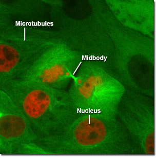
The digital video sequences presented in this section feature epithelial cells undergoing mitosis as visualized through the highly dynamic interactions between fluorescently labeled red histones and green tubulin. Cells enter mitosis through prophase and proceed through division to create a pair of daughter cells. The labeled histones enable visualization of the condensed chromatin as it progresses through various phases, while the tubulin can be seen forming the mitotic spindle as well as the midbodies at telophase.
Video 1 - Run Time: 10 Seconds - An EGFP-tubulin-labeled pig kidney epithelial cell expressing mCherry fluorescent protein fused to histone H2B undergoes mitosis. Choose a playback version: Streaming Video (2.1 MB), Progressive Download (2.1 MB), and MPEG Download (20 MB)
Video 2 - Run Time: 9 Seconds - A pair of daughter cells is created when an EGFP-tubulin-labeled pig kidney epithelial cell expressing mCherry fluorescent protein fused to histone H2B undergoes mitosis. Choose a playback version: Streaming Video (1.7 MB), Progressive Download (1.7 MB), and MPEG Download (17 MB)
Video 3 - Run Time: 16 Seconds - A porcine kidney cell labeled with EGFP-tubulin undergoes mitosis while expressing mCherry fluorescent protein fused to histone H2B. Choose a playback version: Streaming Video (3.2 MB), Progressive Download (3.2 MB), and MPEG Download (29 MB)
Video 4 - Run Time: 16 Seconds - Note the extended midbody as an EGFP-tubulin-labeled pig kidney epithelial cell expressing mCherry fluorescent protein fused to histone H2B undergoes mitosis. Choose a playback version: Streaming Video (3 MB), Progressive Download (3 MB), and MPEG Download (29 MB)
Video 5 - Run Time: 16 Seconds - The chromatids of an LLC-PK1 cell are clearly visible while it undergoes mitosis. Choose a playback version: Streaming Video (4.2 MB), Progressive Download (4.2 MB), and MPEG Download (29 MB)